Both images are acquired simultaneously in a single compression — no additional dose, no additional discomfort. Standard and advanced imaging in one visit.
3D tomosynthesis produces thin-slice images that our radiologists review layer by layer — revealing cancers hidden within overlapping dense tissue that 2D imaging may miss entirely.
Built-in software automatically identifies and marks areas of interest — a second analytical layer that supports our radiologists in delivering a thorough and comprehensive read.
Superior resolution and 3D capability significantly reduce false-positive findings — meaning fewer patients are recalled for imaging that turns out to be unnecessary.
Dense breast tissue can hide cancers on standard 2D mammography. 3D tomosynthesis performs measurably better in dense breasts, detecting more and reducing missed findings.
Every mammogram is personally reported by our team of specialist diagnostic radiologists — not a generalist, not an algorithm. Expert interpretation on every scan.

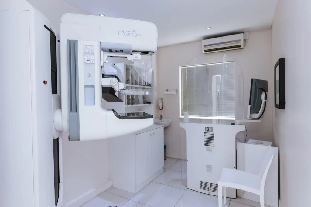
Mammography is the most effective tool for early breast cancer detection. At the Centre of Advanced Medicine, our team of specialist diagnostic radiologists performs and reports all mammograms using the Hologic Selenia Dimensions — a system that acquires both a 2D mammogram and a full 3D tomosynthesis scan in a single compression.
3D tomosynthesis lets our radiologists examine breast tissue one millimetre at a time — detecting cancers hidden in overlapping tissue that a standard 2D image can miss. Fewer callbacks. Earlier detection. Greater confidence in every result.
Screening mammograms are available without a referral. Walk-in bookings are welcome.
No referral is required for screening mammography — walk-in bookings are welcome. A referral is recommended for diagnostic mammograms and required for medical aid purposes.
Current guidelines recommend annual screening from age 40 for women of average risk. Women with a family history of breast cancer or other risk factors may be advised to start earlier — discuss with your GP.
A 2D mammogram produces two flat images per breast. 3D tomosynthesis produces a series of thin-slice images that our radiologists examine layer by layer — particularly valuable in dense breast tissue and significantly reducing false-positive recalls. At the Centre of Advanced Medicine, both are acquired in a single compression.
Compression is necessary for a clear image. Most patients describe it as briefly uncomfortable rather than painful. Avoid scheduling during the week before your period when breasts tend to be more sensitive. Our radiographer will guide you throughout.
Wear a two-piece outfit for easy access. Do not apply deodorant, powder, or cream to the breast or underarm area on the day of your appointment — these can appear on the images. Bring prior mammogram reports if available.
Performed and reported by our team of specialist diagnostic radiologists. 2D and 3D tomosynthesis in a single compression using the gold-standard Hologic Selenia Dimensions. No referral required for screening.
All rights Reserved. Dr Jeff Swartzberg.Designed by FIG Media