An ultrasound scan is a painless, safe procedure that uses high-frequency sound waves — not radiation — to create real-time images of organs, structures, and tissues inside your body. It is one of the most commonly used diagnostic imaging tests available.
How it works:
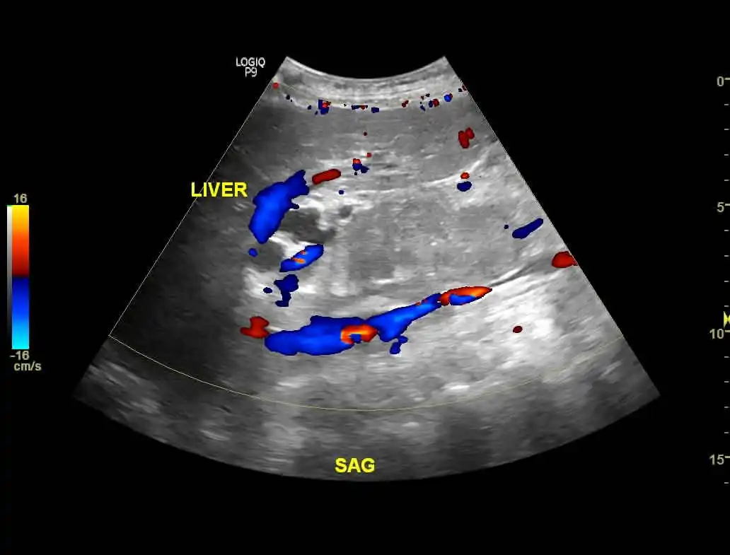
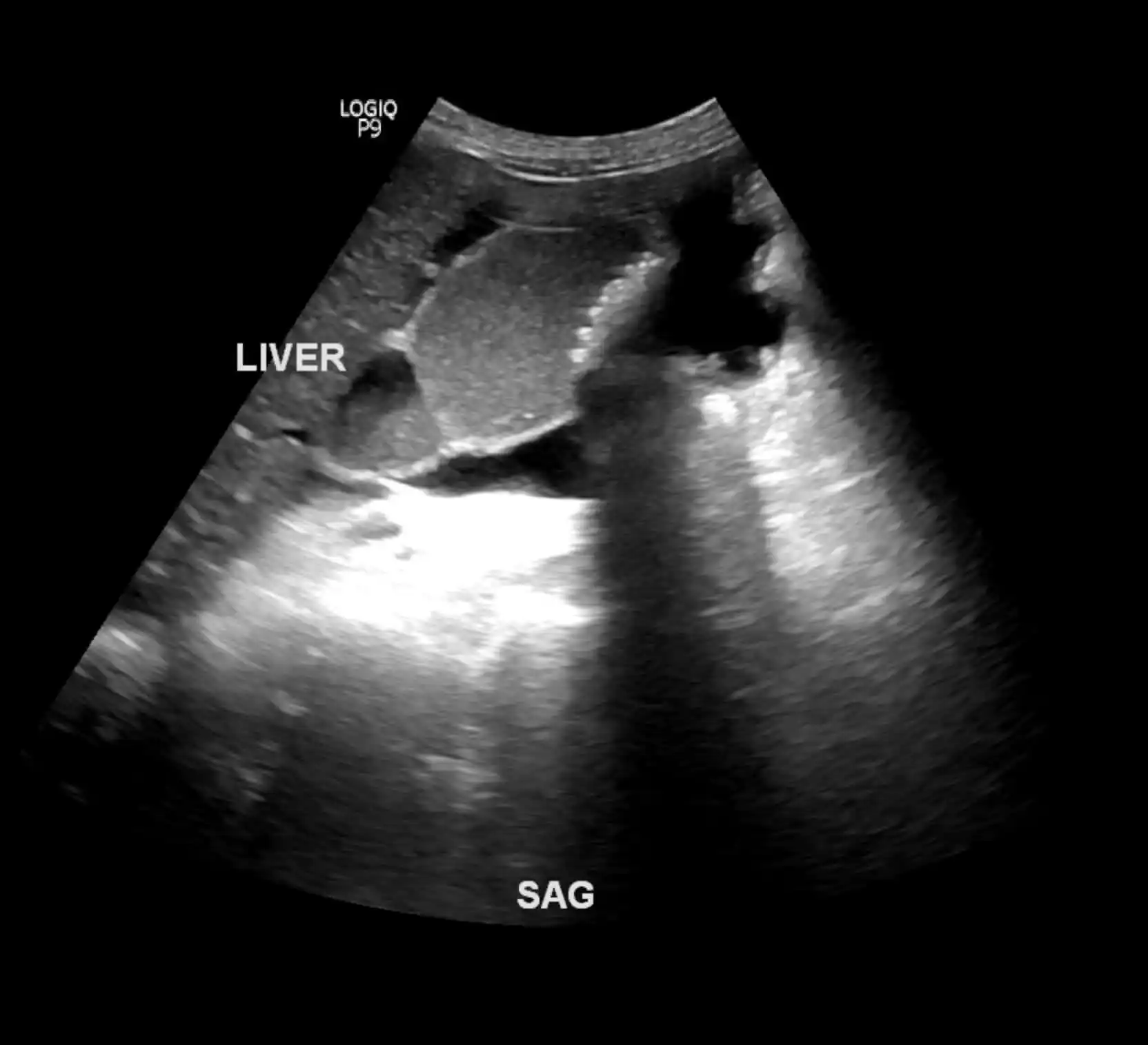
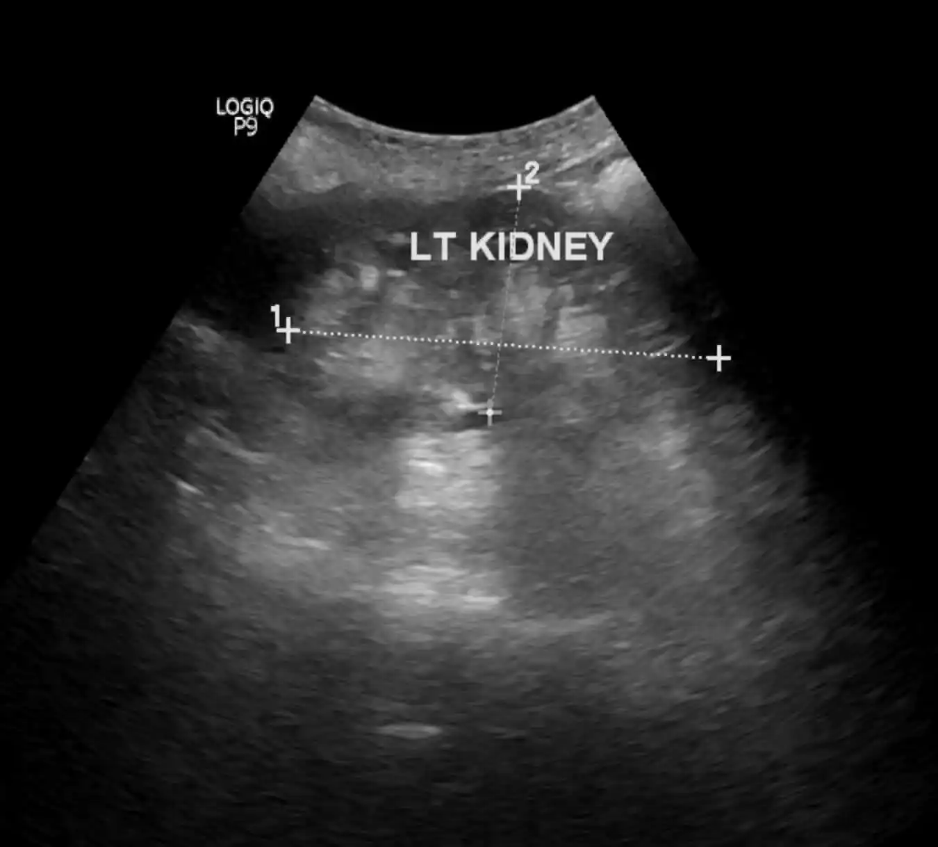
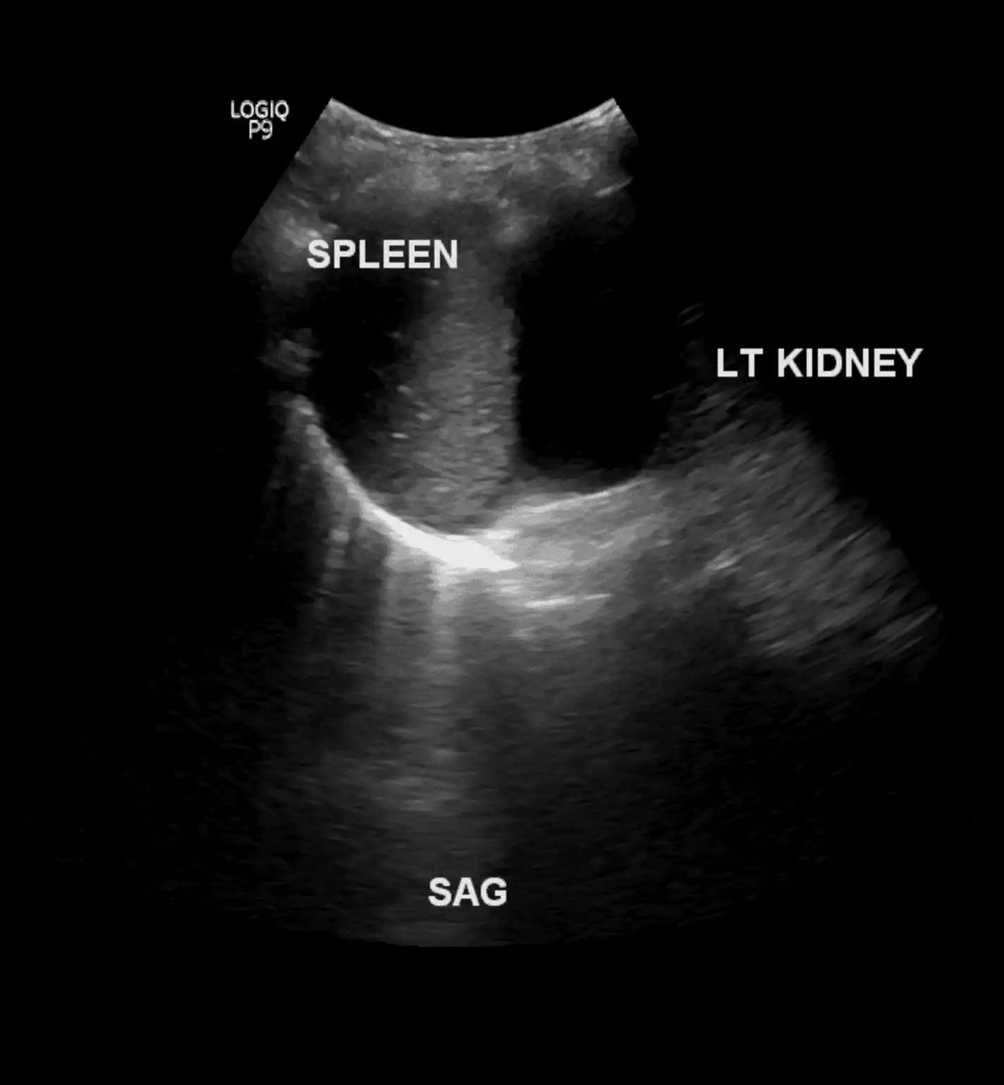
The ultrasound probe sends sound waves into the body, which bounce back as echoes when they hit structures of different densities. These echoes are converted into images displayed on a monitor, allowing our radiologists to assess the size, shape, and consistency of organs and detect abnormalities.
What ultrasound is used for:
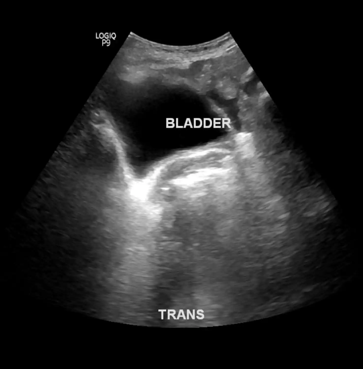
Ultrasound is a versatile diagnostic tool used across many areas of medicine. It is routinely used to monitor fetal growth, development, and screen for abnormalities during pregnancy. In the abdomen, it helps evaluate the liver, gallbladder (including gallstones), pancreas, kidneys, and bladder. For breast and thyroid concerns, ultrasound can distinguish between solid tumours and fluid-filled cysts. It is also used to detect aneurysms and blood vessel abnormalities, as well as to examine the ovaries, testes, and lymph nodes.